SENOLOGICAL RADIOLOGY at J|medical
There are instrumental exams that have become fundamental in the prevention and diagnosis of certain diseases. Among these exams, senological radiology plays a very important role. The doctor may request this specific check from the senologist radiologist.
When is this exam requested? In addition to patients with already diagnosed senological disease, the doctor may request an in-depth study also for women who complain, for example, of the presence of nodules, nipple discharge, inflammatory and/or painful states affecting one or both breasts.
Is J|medical structured to perform these checks? There is a specific area, inside the center, dedicated to senological diagnostics. It is a department that integrates with other services aimed at women, such as gynecology and pelvic rehabilitation.
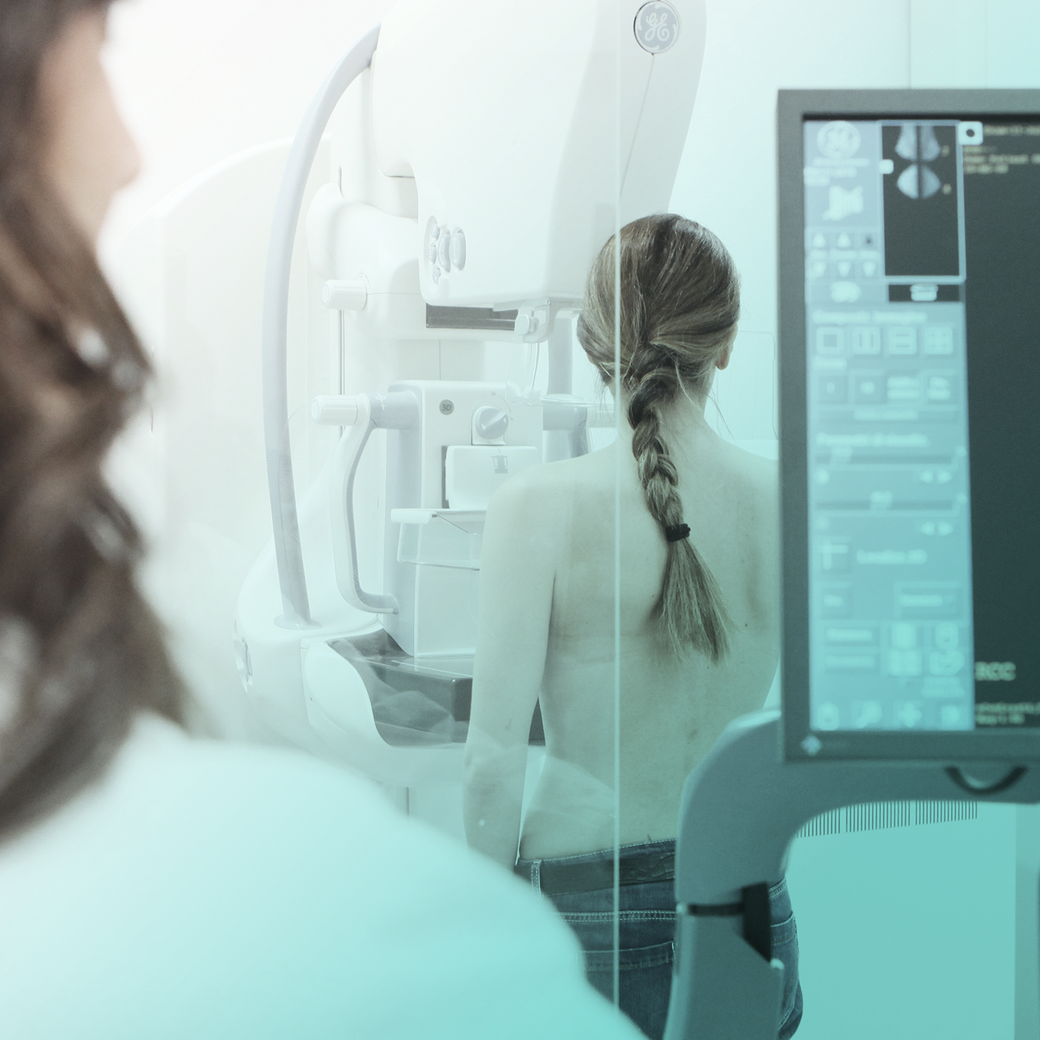
What advantages does this integration bring? It allows women to undergo a complete path that includes mammography, tomosynthesis (3D mammography), ultrasound, and the innovative automated breast ultrasound (ABUS).
So these are both control and prevention exams? Absolutely yes. Senological diagnostic imaging deals with evaluating the conditions of the breast with the main objective of early diagnosis of breast cancer. Today we know that early diagnosis of breast cancer increases the chances of curing and eradicating this disease.
What services do I find at J|medical? The services offered are mainly: Senological visit, 3D mammography (tomosynthesis) and bilateral breast ultrasound Senological visit and breast ultrasound Senological visit and three-dimensional breast ultrasound with Invenia ABUS Do the exams to be done change based on women’s age? Yes. For patients over 40, the ideal path is represented by the senological visit, 3D mammography, and ultrasound. The combination of these methods allows for high diagnostic sensitivity in the search for breast pathologies, with particular reference to the identification of small tumors. For patients under 40, however, the ideal path involves the senological visit and breast ultrasound, even better if performed with 3D ABUS technique. This combination allows for the timely identification of nodules, cystic lesions, and other anomalies.